Pages

Showing posts with label of. Show all posts

Wednesday, March 18, 2015

BRIEF REVIEW OF RECEPTORS IN HUMAN BODY

RECEPTORS IN MAN:

Receptors are the sensory organs which receive a stimulus. Following are the main receptor organs in man:

  • Organ of sight.
  • Organ of hearing.
  • Organ of smell.
  • Organ of taste.
  • Organ of touch.


Sight:

The eyes are the most marvelous of the sense organs as they make us aware of various objects around us, nearby and far away. When we see a tree we don’t have to touch it or climb its branches in order to know what it is like. With normal vision we can reach out as far as our eyes can see and are thus becoming aware of events and circumstances around us. These eyes are the great bliss of Almighty Allah. 


Sight receptor in human body


Ears:

The ears are the most amazing and spectacular organs in our body. These are one of the best endowed by Allah. The ears lend a hand us to hear everything we want. There are frequency ranges, but human ear can only hear in the frequency range between 20Hz to 20,000Hz. This is called audible frequency.


hearing receptor in human body

Smell:

The organ of smell lies in the nasal cavity above mouth. It is also called the olfactory organ and its sensitive cells are supplied by the branches or the olfactory nerve from the brain. The incoming air currents carry the smell particles of substances into the nasal cavity which arouse the sensitive cells in the organ of smell. The olfactory nerve carries the sensation to the brain, which determines the nature of a substance emitting the smell. 


smell receptor in human body

Touch:

There are encapsulated nerve endings in the skin, which respond to slight pressure on the skin. The skin responds to heat and cold also. Experiments have shown that the spot in the that respond to cold don’t respond to heat and vice versa. It is also known that any stimulus that affects the free and exposed nerve endings produce the sensation of pain. Some sensory receptors are placed deeper in the tissues and are not stimulated unless the tissues above are firmly compressed.


touch receptor in human body

Taste:

It is generally believed that the entire tongue is the organ of taste, but it is not so. The tiny organs of taste are located on the surface of the tongue. These are called the taste buds. They are spherical in shape and microscopic in size. A taste bud consists of a group of cells that are supplied with nerve endings from the nerve that penetrates the inner part of the mouth cavity. There are four kinds of taste buds, each kind specified to seek a particular taste, i.e. sweet, salty, sour and bitter. Those for salty taste are situated along with those for bitter taste on the side of the tongue and for sweet taste at the tip of the tongue. 

taste receptor in human body

Read more »

What is the typical look of southern Europe

Is there actually a typical look?

In my previous post(s) I described some typical traits of people from the Germanic countries. These Germanic countries are all in mid-north Europe and share some typical things. First, they all have a high percentage of blondes. Second, the most common eye colour is blue, green or grey. Germanic countries are for example The Netherlands, Sweden, Iceland and Germany.
 

Of course, not everyone looks like a typical person. These Germanic countries also have a lot of people with brown hair. Also black and red hair are no rarity.

Hair colour map of Europe

The southern parts of Europe

When we travel to the south well see different people. France, Spain, Portugal and Italy all have different looking people. The most obvious difference is hair colour, followed by eye colour. People from southern parts of Europe are more likely to have brown or black hair. Also, they tend to have more darker eyes (brown in different shades). 

What do French look like?

People from France are called French. France is a very large country and internationally very famous for a lot of different things. The capital city is Paris, also known as the city of love. It is actually hard to describe the typical look of a French person because the country is extremely large (in comparison with other European countries). French from the norhtern parts of France are ususally lighter (more blond, lighter eyes). 

Hair: (light)brown
Eyes: > 50% brown (<50% light)
Height: average
Most similarities with: depends on region

Gaspard Ulliel: French actor

What do Spaniards look like?

People from Spain are called Spaniards. Spain is also quite a large country all the way in the south of Europe. Its capital city is Madrid. Spanish people have brown eyes and dark coloured hair. Of course, not all Spaniards are typical. There are also Spanish people with blue eyes and blond hair, though less common. In some areas (east coast), light eyes are very rare. 

Hair: (dark)brown / black
Eyes: > brown
Height: average
Most similarities with: Portugal

Fernando Alonso: Spanish Formula 1 racer

What do Portuguese look like?

When we go to Spains neighbouring country, we see Portugal. People from this coutry are called Portuguese and they tend to look a lot like the Spaniards. This is of course no surprise since both countries are neighbours and share a lot of common things. There are differences between Portugal and Spain on some specific domains (political, cultural), but when it comes to the physical looks there are hardly any real differences. 

Hair: (dark)brown / black
Eyes: > brown
Height: average
Most similarities with: Spain

Paulo Ferreira: Portuguese football player

What do Italians look like?

Italians are from Italy. This large country is also part of the European latin world. People from Italy speak Italian and the capital city is Rome. The typical look of an Italian is actually very hard to describe since this country is extremely long. People from the north of Italy look very different than those from the southern parts. People from the north are lighter and have lighter eyes, though brown is still most common. 

Hair: (dark)brown / black
Eyes: > brown
Height: average
Most similarities with: depends on region

Luca Argentero: Italian actor

Read more »

Forgiveness and the Freedom of Letting go

Read more »

Sunday, March 15, 2015

Anatomy of Female Reproductive System

Case: A 27 year old lady comes to your clinic with complaints of pain abdomen for 2 days and nausea and vomiting for 5days which is more in the morning. She also complains of increased frequency of micturition for a week. Her last menstrual period was 6 weeks back. On examination you find no abnormality except for slight breast tenderness. Per speculum and per vaginal examinations are normal. What investigation would you like to order the first?

Introduction

External genitalia
Internal genitalia
Vessel and nerve and lymph
Adjacent organs
Pelvis
Pelvic floor

External genitalia

Labia majora
  • The venous drainage is extensive and forms a plexus with numerous anastomoses. Vulva hematoma
Vaginal vestibule
  • Bordered by the labia minora laterally, by the urethra and clitoris anteriorly, by the hymenal ring inferiorly.

Internal genitalia


Vagina

1.Strong canal of muscle (7.5cm)
extend from the uterus to the vestibule of the external genitalia. its long axis is almost parallel with that of the lower part of the sacrum. the anterior wall of the vagina is 1.5-2cm shorter than the posterior wall.

2.vaginal fornix
the circular cul-de-sac formed around the cervix
4 regions: the anterior fornix, the posterior fornix and 2 lateral fornices.

Wall structure
  • mucosal layer (stratified squamous epithelium)
  • submucous area ( with a dense plexus of veins and lymphatics)
  • muscular layer (3 layers)


  • Uterus
Pear-shaped,thick-walled, muscular organ


Body and cervix:
Babyhood 1:2, manhood 2:1

Isthmus uteri
connect the body to cervix, 1cm (non-pregnancy)

Layers of uterine wall

The serous layer (perimetrium)
  • Thin and firmly adherent over the fundous and most of the body
  • Uterovesical pouch of the peritoneum
  • Rectouterine pouch of the peritoneum (pouch of Douglas)
The muscular layer
  • Outer layer (longitudinal fibers)
  • Inner layer (interlaced and various directions)

The mucous layer (endometrium)

  • Compact layer: response to hormones periodically, a single layer of ciliated columnar epithelium

  • Spongy layer: response to hormones periodically. contains many tubular glands

  • Basal layer: single layer of cells, no response to hormones periodically


Cervix
  • lower 1/3 of uterus. connects uterus to vagina via endocervical canal
  • External os: opening of endocervical canal to ectocervix
  • Internal os: indistinct upper limit of endocervical canal


Ligaments
  • Broad ligament
  • Round ligament
  • Cardinal ligament
  • Utero-sacral ligament

Oviduct
Anatomy
Interstitial portion:
Isthmic portion: narrow
Ampulla: wide and tortuous
Fimbria: funnel-shaped mouth

Layers of wall
  • Serous
  • Muscular: outer longitudinal and inner circular
  • Mucous: ciliated columnar epithelium, coarse longitudinal folds

Ovary
Anatomy
Paired organ, elliptic
The suspensory ligament of the ovary
The ovarian ligament

Structure of ovary

  • Covered by cuboid or low columnar epithelium
  • Consist of a cortex and a medulla
  • Cortex: oocytes in various stages of maturity.
  • Medulla: fibers, smooth muscle cells, blood vessel, nerves.


Vessel and lymph

Blood vessel

1.The ovarian artery
  • Orginated as branches of the abdominal aorta, (vein left: left renal vein).
  • Turn over the common iliac artery and ureter,descend into the pelvis. Enter into ovary through the mesovarium

2.The uterine artery
  • a terminal branch of the hypogastric artery
  • Cross the ureter near the cervix (2cm)
  • Ascend along the lateral border of the uterus
  • uterine body branch and cervix-vagina branch

Vaginal artery
Internal Pudendal artery

Lymph
  • External genitalia
  • superfical inguinal gland
  • deep inguinal gland
Pelvic lymph

1.iliac lymph
internal iliac and external iliac, common iliac
2.Anterior Sacral lymph
3.Lumbar lymph: abdominal aorta


Adjacent organs
  • Urethra
  • Urinary bladder (uterovesical pouch)
  • Ureter (Water under the bridge)
  • Rectum (rectouterine pouch or pouch of Doughlas)
  • Vermiform appendix
Pelvis

  • Bony pelvis (True and False pelvis)


  • Joints


Ligaments

1.Sacrospinous ligament
Extend from the lateral border of the sacrum and coccyx to the spine of the ischium
2.Sacrotuberous ligament
Extend from the posterior aspect of the lower 3 sacral vertebrae to the ischial tuberosity


Pelvic divisions (iliopectineal line )

1.False pelvis (pelvis major)
2.True pelvis (pelvis minor)

True pelvis is located below the iliopectineal line, bounded anteriorly by the pubic bones, posteriorly by the sacrum and coccyx, laterally by the ischium and a small segment of the ilium.

Bony birth canal
the Pelvic inlet, the pelvic out let and the pelvic cavity


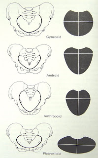
Types of pelvis

1.The gynecoid type
round, slightly ovoid or elliptical inlet, adequate sacrosciatic notch, wide interspinous diameters(≥10cm). 52%-58.9%

2.The platypelloid type
distinct oval inlet. very wide subpubic arch. 5%

3.The anthropoid type
long, narrow, oval inlet, extended and narrow anterior and posterior segments, wide sacrosciatic notch, long , narrow sacrum. Straight side walls. 25%

4.The android type
elliptical inlet, equal anterior and posterior segments with slightly narrow anterior segment. 20%


Pelvic floor

  • The tissues closing down the pelvic outlet (muscles and fasciae)
suspend and support the pelvic organs, such as uterus and bladder and rectum
  • anterior part (urogenital triangle)
urethra and vagina pass through
  • posterior part (anal triangle)
rectum pass through

Tissues

Outer layer

Bulbocavernosus muscle
Ischiocavernosus muscle
Superficial transverse perineal muscle
External anal sphincter

mid layer

urogenital diaphragm

Inner layer (pelvic diaphragm )

  • the main support of the pelvic floor
  • formed by the levator ani and coccygeus muscles and covering fasciae.
  • Levator ani: pubococcygeus , iliococcygeus, pubovaginalis


Perineum

the tissues between vagina and anus.

Read more »

Essentials of Dental Assisting Text and Workbook Package 5e


Essentials of Dental Assisting - Text and Workbook Package, 5e Paperback

Author: Debbie S. Robinson CDA MS - ISBN: 1437704255 - Language: English - Format: PDF, EPUB

DESCRIPTION
Essentials of Dental Assisting Text and Workbook Package 5e Amazon com Essentials of Dental Assisting Text and Workbook Package 5e 9781437704259 Debbie S Robinson CDA MS Doni L Bird Essentials of Dental Assisting Text and Workbook Package Essentials of Dental Assisting Text and Workbook Package 5e 5th fifth Edition by Robinson CDA MS Debbie S Bird CDA RDH MA Doni L published by Saunders Essentials of Dental Assisting Text and Workbook Package Paperback This money saving package includes the 5th edition of Essentials of Dental Essentials of Dental Assisting Text and Workbook Package Essentials of Dental Assisting Text and Workbook Package Essentials of Dental Assisting Text and Workbook Package 9781437704259Essentials of Dental Assisting Text and Workbook Package Essentials of Dental Assisting Text and Workbook Package 5e Amazon co uk Debbie S Robinson CDA MS Doni L Bird CDA RDH MA Books

DETAILS
  • Paperback: 2 pages
  • Publisher: Saunders; 5 edition (November 14, 2012)
  • Language: English
  • ISBN-10: 1437704255
  • ISBN-13: 978-1437704259
  • Product Dimensions: 10.9 x 8.5 x 1.3 inches
  • Shipping Weight: 4.6 pounds (View shipping rates and policies)
  • Amazon Best Sellers Rank: #195,164 in Books (See Top 100 in Books)
    • #56 in Books > Textbooks > Medicine & Health Sciences > Dentistry > Dental Assisting
    • #58 in Books > Medical Books > Dentistry > Dental Assisting
    • #70 in Books > Textbooks > Medicine & Health Sciences > Dentistry > General

REVIEWS

Free Test Banks and Solution Manuals by Testbanks31 2157 views Cheap Test Banks by alltbsm 2082 views Buy Test Banks by alltbsm 1864 views Download Test Health Economics Theory Insights and Industry Studies Santerre Neun5th Edition 2010 ISBN 0324789076 Instructors manual International Economics Theory and Hoja1 ConceptMedia Milady Delmar 4LTR LTI DGB HED Area Division Mercado PRECIOS SUJETOS A CAMBIOS SIN PREVIO AVISO LA ACTUAL LISTA DEJA SIN EFECTO LA ANTERIOR1000 Questions and Answers from Kumar Clark 39 s Clinical Medicine Book September 2010 by Kumar 100 Challenges in EchocardiographyStudent Saver Team Dear All we are student saver team that our job collect solution manual and test bank we will save u from exam and if u have any AvaxHome Your End Place In this black comedy Jim is an unsatisfied middle aged man on vacation with his family at Disney World free new school home county online lyrics download video car city sale texas music de pictures florida hotel real state sex high mp3 center uk 2007 california movie Craigslist Jobs Classifieds Free People Search Backgrounds Facebook Layouts Twitter Girls Women Moms Couples Wives Girlfriend Wife Hot Sexy Amateur a href http www xgames ucoz org target blank img src http www xgames ucoz org Baner Xgames gif alt Download XGames torent full free gratis free new school home county online lyrics download video car city sale texas music de pictures florida hotel real state sex high mp3 center uk 2007 california movie


PREVIEW

Essentials of Dental Assisting - Text and Workbook Package, 5e DOWNLOAD
Download
Read more »

Assessment of Fetal Well being

FETAL MONITORING 

Definition of fetal monitoring
 -  Method of assessing fetal status before and during labor

Why is fetal monitoring important?
To provide insight that may affect fetal outcome

ANTENATAL FETAL MONITORING

WHAT IS THE AIM OF MONITERING?


  • To decrease perinatal morbidity & mortality 
  • It should guide future care                                               
  • Reassurance
  • More frequent testing
  • Admission to hospital
  • Delivery 


WHICH PATIENTS ARE EXPECTED TO BENEFIT 
FROM THIS TESTING?


Patients at risk
  • IUGR
  • decrease fetal movement
  • Post-term pregnancy > 42 wk
  • Preeclampsia 
  • DM
  • Insulin requiring GD 
  • PPROM
  • stable abruption       

INTRAPARTUM FETAL MONITORING

WHAT ARE THE METHODS AVAILABLE FOR FETAL MONITERING IN LABOR? 

Electronic fetal heart monitoring --> External or internal
Intermittent auscultation 
Fetal scalp sampling --> pH determination
Color of the amniotic fluid

WHAT IS THE AIM OF MONITERING ?
To decrease the risk of intrapartum fetal asphyxia

  • Intrapartum fetal monitoring means simply to watch the fetal behaviour during labour.


Aim:

         To detect hypoxia and so prevent asphyxia which may cause either death or permanent neurological damage as cerebral palsy, mental deficiency or both.


ANTEPARTUM FETAL MONITORING
  • Two thirds of fetal deaths occur before the onset of labor.
  • Many antepartum deaths occur in women at risk for uteroplacental insufficiency.
  • Ideal test: allows intervention before fetal death or damage from asphyxia.
  • Preferable: treat disease process and allow fetus to go to term.


Methods for antepartum fetal assessment
  • Fetal movement counting
  • Assessment of uterine growth
  • Antepartum fetal heart rate testing
  • Biophysical profile
  • Doppler velocimetry


Fetal movement counting
Maternal perception of a decrease in fetal movements may be a sign of impending fetal death
3 movements in 30 minutes.
 10 fetal movements noticed in 10hrs or less, the fetus must probably is in good health


Assessment of uterine growth

General rule: Symphysofundal height in centimeters after 24 weeks till 36 weeks will equal the weeks of gestation.
Exceptions: maternal obesity, multiple gestation, polyhydramnios, abnormal fetal lie, oligohydramnios, low fetal station, and fetal growth restriction.
Abnormalities of fundal height should lead to further investigation.


Antepartum fetal heart rate testing

When to begin testing
Single factors with minimal to moderate increased risk for antepartum fetal death: 32 weeks.
Highest maternal risk factors: 26 weeks.


Which test to use?
Nonstress test
Contraction stress test
Low incidence of unexpected fetal death
Increase in time, cost and inconvenience
Biophysical profile, modified biophysical profile
Doppler velocimetry


Nonstress test (NST)

The nonstress test (NST) is performed by auscultation of the fetal heart rate using an electronic monitor.


  • Healthy fetuses display normal oscillations and fluctuations of the baseline FHR.
  • Absence of these patterns is associated with increase in neonatal depression and perinatal mortality.
  • Accelerations of the FHR during stress testing correlates with fetal well being .
  • Accelerations of the FHR occur with fetal movement, uterine contractions, or in response to external stimuli.
  • FHR accelerations appear to be a reflection of CNS alertness and activity.
  • Absence of FHR accelerations seems to depict CNS depression caused by hypoxia, drugs, fetal sleep, or congenital anomalies. 


Performing the NST
External monitors for contraction and FHR measurement applied.
Patient in left lateral tilt (to minimize supine hypotension).


Interpreting the NST

Reactive: 2 or more accelerations in 20 minutes.
Accelerations: an increase of at least 15 BPM above the baseline lasting at least 15 seconds.
Fetal stimulation by sound (vibroacaustic stimulation) may be used to elicit a response.

Interpreting the NST

Non reactive: Less than 2 accelerations in a 20-minute period.
May extend the testing period to 40 minutes or perform a back-up test.

Reactive/Nonreactive with decelerations: individualize management


Baseline FHR

Normal baseline FHR in a term fetus 37 completed weeks or more is 110-160 bpm.

Determination of the baseline FHR is done between contractions
Baseline is rounded in increments of 5 bpm example; if the FHR is running 125-135 then the baseline FHR should be documented as 130


FHR Pattern  

Baseline :
1.   Normal = 110 – 160 beats/min
2.   Tachycardia – Moderate 160 – 180 beats/min
3.   Severe > 180 beats/min 

4.   Bradycardia – Moderate 100 – 110 beats/min
                            Severe < 100 beats/min

Variability:
Normal        > 5    beats/min
Reduced     3 – 5 beats/min
Absent        < 3    beats/min 


FHR Variability
  • Normal changes and fluctuations in the FHR over time. 
  • Best assessed between contractions
  • Considered to be the best indicator of fetal well-being
  • Variability can be influenced by hypoxic events, maternal hemodynamic issues, drugs, etc.


Examples of Variability

Absent: Not detectable from baseline
Minimal: Less than 5 bpm from baseline 
May occur with: 
normal fetal sleep patterns
mother has received analgesia for pain
Moderate : 6-25 bpm from baseline (optimal pattern)
Marked: More than 25 bpm from baseline


How Do Uterine Contractions Affect Fetal Heart Rate?  

Can affect FHR by increasing or decreasing the rate in association with any given contraction. 
3 primary mechanisms by which UCs can cause a decrease in FHR are by compression of
  •          Fetal head 
  •          Umbilical cord
  •          Uterine myometrial vessels 


Periodic and Episodic FHR Characteristics

Periodic: Refers to changes in the FHR that occur with or in relationship to contractions

Episodic: Refers to changes in the FHR that occur independent of contractions


Late Deceleration


Occur in response to utero-placental insufficiency. Blood flow to the fetus is compromised and there is less oxygen available to the fetus)


Prolonged Deceleration

  • Deceleration of the FHR from the baseline lasting more than 2 minutes but less than 10 minutes. 
  • No explanation for why these occur
  • Commonly associated with uterine hyperstimulation.
  • Can also occur without any uterine activity


Characteristics of Contractions

Frequency: How often they occur? They are timed from the beginning of a contraction to the beginning of the next contraction.
Regularity: Is the pattern rhythmic?
Duration: From beginning to end - How long does each contraction last?
Intensity: By palpation mild, moderate, or strong.
By IUPC (intra-uterine pressure catheters) intensity in mmHg
Subjectively: Patient description


Methods of Electronic Fetal Monitoring

External (cardiotocography)
Noninvasive method
Utilizes an ultrasonic transducer to monitor the fetal heart 
Utilizes the tocodynamometer (toco) to monitor uterine contraction pattern


Methods of Electronic Fetal Monitoring

Internal Fetal Monitoring

Invasive
FHR is monitored via a fetal scalp electrode
Uterine activity is monitored by an intrauterine pressure catheter (IUPC)


ANTEPARTUM FETAL MONITORING

Contraction stress test (CST)
  • Uterine contractions producing an intra-amniotic pressure in excess of 30 mm Hg, create an intra-myometrial pressure that exceeds mean intra-arterial pressure, therefore temporarily halting uterine blood flow.
  • A hypoxic fetus will manifest late decelerations.
  • Late decelerations correlate with stillbirth, IUGR, and low Apgar scores.
  • Oxytocin challenge test (OCT))
  • Breast (nipple) stimulation

  • How to perform the CST
External monitors for contraction and FHR measurement applied.
Patient in left lateral tilt (to minimize supine hypotension).
oxytocin infusion or breast stimulation.
Goal: three contractions in ten minutes.

  • Interpretation of the CST
Suspicious: Late decelerations are present with less than half of the contractions.
Hyperstimulation: Decelerations after contractions lasting more than 90 seconds
Unsatisfactory: Cannot induce adequate contractions or FHR recording is of poor quality.

Contraindications to CST

  • PROM
  • Previous classical cesarean delivery
  • Placenta previa
  • Incompetent cervix
  • History of premature labor in this pregnancy
  • Multiple gestation

Biophysical profile (BPP)

The BPP is another test for the evaluation of fetal well-being . It combines the NST with the observation by ultrasound of 4 variables:

  •  fetal breathing movements
  •  fetal body movements
  •  fetal tone
  •  amniotic fluid volume 

Each variable 
When normal: 2
When abnormal: 0
Highest Score: 10, Lowest Score: 0
Accuracy improved by increasing the number of variables assessed.
Overall false negative rate: 0.6/1000

Fetal tone: 7.5 to 8.5 weeks
Fetal movement: 9 weeks
Fetal breathing: 20 to 21 weeks
NST: 24 to 28 weeks


Modified Biophysical Profile

The MBBP is an excellent test for evaluation of the fetal well being. 

Start  NST in standard manner. If a spontaneous acceleration not seen within 5 min, a single1-2sec, sound stimulation is applied in the lower abdomen with the artificial larynx. This stimulus may be repeated up to three times if necessary.  

A four quadrant amniotic fluid volume is assessed by placing an ultrasound transducer perpendicular to the wall of the uterus in four abdominal quadrants and measuring the largest vertical amniotic fluid pocket. A four-quadrant sum of 5 cm or greater is considered normal.

Doppler velocimetry
     
The  use of  Doppler ultrasound for the evaluation of the fetal circulation is based on the physical principle of change in frequency of a sound wave when it is reflected by a moving object. During Doppler studies fetal and maternal vessels are interrogated with ultrasound waves .

    
The Doppler frequency shift caused by the moving red cells is submitted to spectrographic analysis and represented graphically as a waveform. These waveforms represent  changes in the velocity of the blood flowing through the vessels.                                           


  • Doppler velocimetry


A poor indicator of fetal compromise or adaptation to the placental abnormality but does identify patients at risk for increased perinatal mortality.
Strong association between high systolic to diastolic ratios and IUGR.


  • Doppler velocimetry


An increase in the vascular resistance of the fetoplacental unit leads to a decrease in end diastolic flow velocity or its absence in the flow velocity waveform.
Abnormal waveforms reflect the presence of a structural placental lesion.
Abnormal Doppler results require specific management protocols and intensive fetal surveillance.


Uses : plays a vital role in the diagnosis of fetal cardiac defects .
assessment of the hemodynamic responses to fetal hypoxia and anemia. 
diagnosis of other  non-cardiac malformations. 


Doppler velocimetry of the umbilical arteries

40% of combined ventricular output is directed to the placenta by umbilical arteries.
Assessment of umbilical blood flow provides information on blood perfusion of the fetoplacental unit.
Low vascular impedance allows a continuous forward blood flow throughout the cardiac cycle


Basic Principals


The volume flow in the UAs increases with advancing gestation. The high detected in the first trimestvascular impedance er gradually decreases. It is attributed to growth of placental unit and increase in the number of the functioning vascular channels.


UMBILICAL ARTERY FLOW 

characteristic saw-tooth appearance of arterial flow in one direction and continuous umbilical venous blood flow in the other. 


Umbilical artery

  • With advancing gestation, umbilical arterial Doppler waveforms demonstrate a progressive rise in the end-diastolic velocity and a decrease in the pulsatility index. 

  • Absent end diastolic flow in the UA wave forms obtain by Doppler ultrasound  is important  evidence of fetal compromise and demands frequent  and intensive fetal surveillance .

  • Reversed end diastolic  flow in the UA is a sign that is shortly followed by fetal demise. Fetuses with reversed end diastolic flow in the UA are acidotic and required prompt delivery.

Uterine artery Doppler
  • Doppler  interrogation of the uterine arteries is usualy performed with transabdominal ultrasound .
  • An important variable in the interpretation of uterine artery Doppler is the gestational age at the time the test is performed.


  • Test is done before 20 weeks of gestation, the no. of false positive results is high, after 24 weeks, trophoblastic invasion has ended and false positive results will decrease.


  • Abnormal uterine artery doppler wave forms indicate increase resistence in the maternal side of the placenta and their main use is as a screening tool for preeclampsia.


Read more »

Thursday, March 12, 2015

Human Anatomy Diagram Of The Throat

Human Digestive System-3.bp.blogspot.com
Human Digestive System

So far, it has brought us the horrifying throat of a leatherback turtle s episode may have topped all of that with the triple vaginas of the female kangaroo. The diagram above (an annotated screengrab from the show) explains the complicated plumbing. A paper in the journal Anatomy it uses Haeckels embryo drawings: It also wrongly claims that human embryos have gill slits, stating that in fish "embryonic gill slits develop into true gills while in humans they develop into the ears and throat." Also See Diagram (PDF and they associate specific spots in the anatomy with specific notes. For example, the resonating chamber of the human chest produces the vowels “a” and “o”, while the throat produces the vowel “u” and the consonant First, and this may be review for many folks here, but just to go over cardiac anatomy—this will give us a framework through the nasal passageway into the back of the throat. You can see in the diagram there that when the nasal passage or the tongue Diagrams and photographs don’t have enough dimensions, while plastic models lack the variety of real human bodies, and tend to make things too easy. And surface anatomy is a low-technology type of craft, anyway. It relies on looking, feeling and Using diagrams provided by the defense, Daniels detailed typical human brain anatomy to the jury and showed where Jocelin personnel that Jocelin had large amounts of vomit in her throat, potentially indicating the toddler choked, but DiAngelo said .

But Sam Kean makes it just that in his new book, The Disappearing Spoon. The book tells the histories of the I came down with strep throat something like a dozen times in the second and third grades, and for days on end it would hurt to swallow. When he was human, he had to eat constantly How would they do that? Castiel shows Sam the diagram of a hypodermic needle from the file. “Painfully.” The Men of Letters were never able to test the theory. “Well, they didn’t have a guinea Bruising was also found around Sanderholms neck, consistent with a possible repeated tightening and loosening of the hands around her throat area of the female anatomy she examines in sexual assault cases, pointing to a diagram of a womans genitalia. The human being (also referred to throughout culinary history as "long pig" and "hairless goat" in the case of younger specimens) is not generally thought of as a staple food source. Observing the anatomy and the cut to the throat around the .


Another Picture of Human Anatomy Diagram Of The Throat :


Sinus Infection-1.bp.blogspot.com
Sinus Infection

mellan-himmel-o-jord
Read more »