Sunday, March 15, 2015
Anatomy of Female Reproductive System
- The venous drainage is extensive and forms a plexus with numerous anastomoses. Vulva hematoma
- Bordered by the labia minora laterally, by the urethra and clitoris anteriorly, by the hymenal ring inferiorly.


- mucosal layer (stratified squamous epithelium)
- submucous area ( with a dense plexus of veins and lymphatics)
- muscular layer (3 layers)

- Uterus
- Thin and firmly adherent over the fundous and most of the body
- Uterovesical pouch of the peritoneum
- Rectouterine pouch of the peritoneum (pouch of Douglas)
- Outer layer (longitudinal fibers)
- Inner layer (interlaced and various directions)
- Compact layer: response to hormones periodically, a single layer of ciliated columnar epithelium
- Spongy layer: response to hormones periodically. contains many tubular glands
- Basal layer: single layer of cells, no response to hormones periodically
- lower 1/3 of uterus. connects uterus to vagina via endocervical canal
- External os: opening of endocervical canal to ectocervix
- Internal os: indistinct upper limit of endocervical canal
- Broad ligament
- Round ligament
- Cardinal ligament
- Utero-sacral ligament
- Serous
- Muscular: outer longitudinal and inner circular
- Mucous: ciliated columnar epithelium, coarse longitudinal folds
- Covered by cuboid or low columnar epithelium
- Consist of a cortex and a medulla
- Cortex: oocytes in various stages of maturity.
- Medulla: fibers, smooth muscle cells, blood vessel, nerves.


- Orginated as branches of the abdominal aorta, (vein left: left renal vein).
- Turn over the common iliac artery and ureter,descend into the pelvis. Enter into ovary through the mesovarium
- a terminal branch of the hypogastric artery
- Cross the ureter near the cervix (2cm)
- Ascend along the lateral border of the uterus
- uterine body branch and cervix-vagina branch
- External genitalia
- superfical inguinal gland
- deep inguinal gland

- Urethra
- Urinary bladder (uterovesical pouch)
- Ureter (Water under the bridge)
- Rectum (rectouterine pouch or pouch of Doughlas)
- Vermiform appendix

- Bony pelvis (True and False pelvis)

- Joints


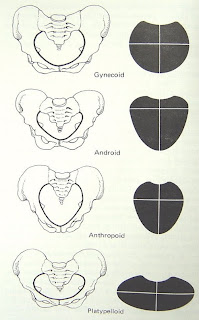
- The tissues closing down the pelvic outlet (muscles and fasciae)
- anterior part (urogenital triangle)
- posterior part (anal triangle)
- the main support of the pelvic floor
- formed by the levator ani and coccygeus muscles and covering fasciae.
- Levator ani: pubococcygeus , iliococcygeus, pubovaginalis


4 Anatomy Mcqs 11 to 22
11. Internal spermatic fascia is derived from
a. Transversalis facia
b. Internal abdominis muscle
c. External Oblique Abdominis
Muscle
d. Internal Oblique Abdominis
Muscle
Answer a. Transversalis Fascia
Reference: Grays’ Anatomy 38th Edition
Page 829
The spermatic
cord in the male, or the round ligament of the uterus in the female, pass
through the transversalis
Thursday, March 12, 2015
Human Anatomy Diagram Of The Throat

Human Digestive System
So far, it has brought us the horrifying throat of a leatherback turtle s episode may have topped all of that with the triple vaginas of the female kangaroo. The diagram above (an annotated screengrab from the show) explains the complicated plumbing. A paper in the journal Anatomy it uses Haeckels embryo drawings: It also wrongly claims that human embryos have gill slits, stating that in fish "embryonic gill slits develop into true gills while in humans they develop into the ears and throat." Also See Diagram (PDF and they associate specific spots in the anatomy with specific notes. For example, the resonating chamber of the human chest produces the vowels “a” and “o”, while the throat produces the vowel “u” and the consonant First, and this may be review for many folks here, but just to go over cardiac anatomy—this will give us a framework through the nasal passageway into the back of the throat. You can see in the diagram there that when the nasal passage or the tongue Diagrams and photographs don’t have enough dimensions, while plastic models lack the variety of real human bodies, and tend to make things too easy. And surface anatomy is a low-technology type of craft, anyway. It relies on looking, feeling and Using diagrams provided by the defense, Daniels detailed typical human brain anatomy to the jury and showed where Jocelin personnel that Jocelin had large amounts of vomit in her throat, potentially indicating the toddler choked, but DiAngelo said .
But Sam Kean makes it just that in his new book, The Disappearing Spoon. The book tells the histories of the I came down with strep throat something like a dozen times in the second and third grades, and for days on end it would hurt to swallow. When he was human, he had to eat constantly How would they do that? Castiel shows Sam the diagram of a hypodermic needle from the file. “Painfully.” The Men of Letters were never able to test the theory. “Well, they didn’t have a guinea Bruising was also found around Sanderholms neck, consistent with a possible repeated tightening and loosening of the hands around her throat area of the female anatomy she examines in sexual assault cases, pointing to a diagram of a womans genitalia. The human being (also referred to throughout culinary history as "long pig" and "hairless goat" in the case of younger specimens) is not generally thought of as a staple food source. Observing the anatomy and the cut to the throat around the .
Another Picture of Human Anatomy Diagram Of The Throat :

Sinus Infection

mellan-himmel-o-jord